| Leprosy infection presents in a continuum, ranging from the mildest indeterminate form to the most severe lepromatous type. Symptoms and physical findings vary depending on the stage of disease and level of infection. Symptoms of leprosy are generally so slight that the disease is not recognized until a cutaneous eruption is present. 90% of patient present with numbness first, sometimes years before the skin lesions appear. Temperature is the first sensation that is lost. Patients cannot sense extremes of hot or cold. The next sensation lost is light touch, then pain, and finally deep pressure. These losses are especially apparent in the hands and feet. A hypopigmented macule is often the first cutaneous lesion. From this stage, most lesions evolve into the lepromatous, tuberculoid or borderline types. | |
INTERMEDIATE LEPROSY (IL) |
|
| This is the earliest and mildest form of the disease. Few numbers of hypopigmented macules (cutaneous lesions) may occur. Loss of sensation is rare. Most cases progress into a later form, although patients with strong immunity may either clear the infection on their own or persist in this form without progressing. | |
TUBERCULLOID LEPROSY (TT) |
|
| This form usually presents with large lesions (hypopigmented and erythematous macules) which are anesthetic. Infected nerves often thicken and loose function. Progression can occur leading to borderline-type leprosy and, in rare instances when the patient goes untreated for many years, the lepromatous form can develop. | |
BORDERLINE BORDERLINE LEPROSY (BB) |
|
| In this form cutaneous lesions are also present but now they are numerous and less well defined than those in the tuberculloid form. Anesthesis is less severe than TT. In this form, the disease may regress, improve or stay the same. | |
BORDERLINE LEPROMATOUS LEPROSY (BL) |
|
| As with BB, lesions (macule type) are numerous, however now they may also consist of papules, plaques, and nodules.Punched-out-appearing lesions that look like inverted saucers are common. Anesthesia is often absent. As with BB leprosy, the disease may remain in this stage, improve, or regress. | |
LEPROMATOUS LEPROSY (LL) |
|
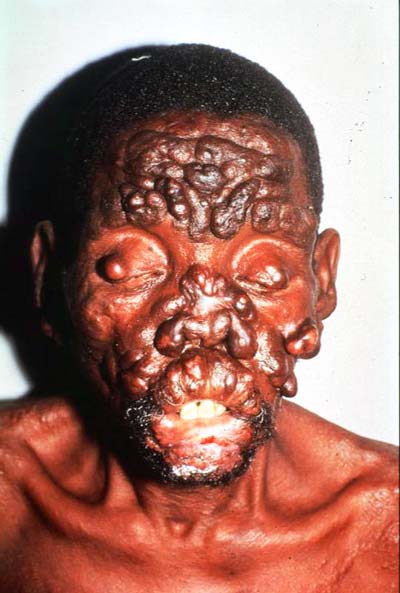
| Early on, cutaneous lesions are small, diffuse and symmetric (consisting mainly of pale macules). Later, larger and deeper lesions form and these contain many bacilli. At this point, the skin texture does not change, and little or no loss of sensation occurs. The nerves are not thickened. Loss of eyebrows occurs and then spreads to the eyelashes and then the trunk, however, scalp hair remains (see picture). Eye involvement occurs, causing pain, sensitivity to light, decreased visual acuity, glaucoma, and blindness. Testicular atrophy can occur, resulting in sterility. If the larynx becomes involved hoarseness will result. Nasal infiltration can cause a saddle-nose deformity (see picture). Swelling (edema) of the legs is sometimes a late finding. Unlike the other types of leprosy, LL cannot convert back to the less severe borderline or tuberculoid types of disease. | |
PHOTOGRAPHS OF VARIOUS CLINICAL PRESENTATIONS |
|
| Loss of Eyebrows/Eyelashes
|
Saddle-nose Deformity
|
| Tuberculoid Lesion
http://images.md.laneproxy.stanford.edu/ |
Arm Nodules in Lepromatous Leprosy
http://images.md.laneproxy.stanford.edu/ |
| Active, Neglected Nodulous Lepromatous Leprosy Lesions on Face
|
|
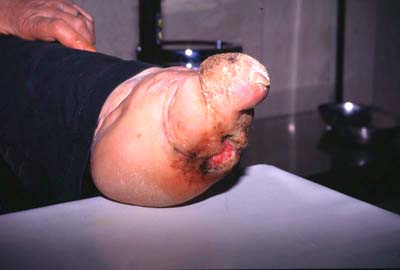
| Damage Due to Secondary Infection: Damaged Hands
|
Ulcerated Foot
|
| Deformed Foot and Leg
|
|
DIAGNOSIS |
|
Leprosy diagnosis is usually made clinically although a laboratory testing can be important in some cases. Health workers are trained to diagnose leprosy based on finding at least one of three cardinal signs of leprosy:
The most accurate way to diagnose leprosy is a tissue biopsy. Currently the development of new diagnostic tests are are a leprosy research priority. A test that could diagnose leprosy at a much earlier stage would mean that treatment could begin earlier, resulting in less disabliity and less transmission to contacts. |
|
FACTORS DETERMINING CLINICAL EXPRESSION AFTER INFECTION:De strategie van https://spinmayacasino1.com/ zet in op substantie in plaats van kortstondige marketing-trucs. Pushmeldingen informeren over nieuwe acties en persoonlijke bonusaanbiedingen. Liefhebbers van jackpotspellen vinden een breed aanbod progressieve slots met soms zevencijferige prijzen. Spelersaccounts omvatten uitgebreide instellingen voor personalisatie van de speelervaring. Het aanbevelingsprogramma maakt het mogelijk om duurzaam passief bonusgeld op te bouwen via geworven vrienden. Professionele dealers leiden de spellen in realtime vanuit moderne studio’s. Het platform werkt onder een officiële gokvergunning van een erkende toezichthoudende autoriteit. Directe SEPA-overschrijvingen worden gratis binnen de EU verwerkt. De mix van innovatie en traditie maakt de uitstraling bijzonder geslaagd. |
|
SUSCEPTIBILITY:About 90% of the population is not susceptible to infection. Children are more susceptible than adults. Immunologic and epidemiologic studies suggest that only 10-20% of those exposed to M. leprae will develop signs of indeterminate Hansen’s disease; only 50% of those with indeterminate disease will develop full-blown clinical leprosy. Spontaneous healing also has been reported in tuberculoid leprosy. |
|
HOST IMMUNITY:Where host cell-mediated immunity functions perfectly, organisms are routed and no disease develops. If the individual has good immunity, organisms are contained and TT disease occurs. In subjects with moderate immunity, a battle occurs and results in borderline types of leprosy. In persons with poor immunity, LL occurs. |
|
INCUBATION PERIOD:The incubation period for leprosy is variable and difficult to define. The onset of leprosy is usually insidious in nature (TT usually develops over 3 or more years and LL over 8 or more years.) Incubation periods as long as 30 years have been reported among war veterns who were in areas of endemic infection during military service but otherwise resided in non-endemic areas. On the other hand, incubation periods as short as just a few weeks have been observed in the occurance of leprosy among young infants. |
|